The list of landmark tools in the fast-developing world of diagnostic imaging goes on, and one such tool is endocavity ultrasound. It provides highly tailored views of many diseases in modern medicine. Of advanced form, ECUS delivers clinical clinicians clear images of internal anatomy and, as such, allows for an improvement both in diagnosis and patient outcome results. An overview on endocavity ultrasound in diagnostic imaging: role, applications, advantages, limitations, and future directions.
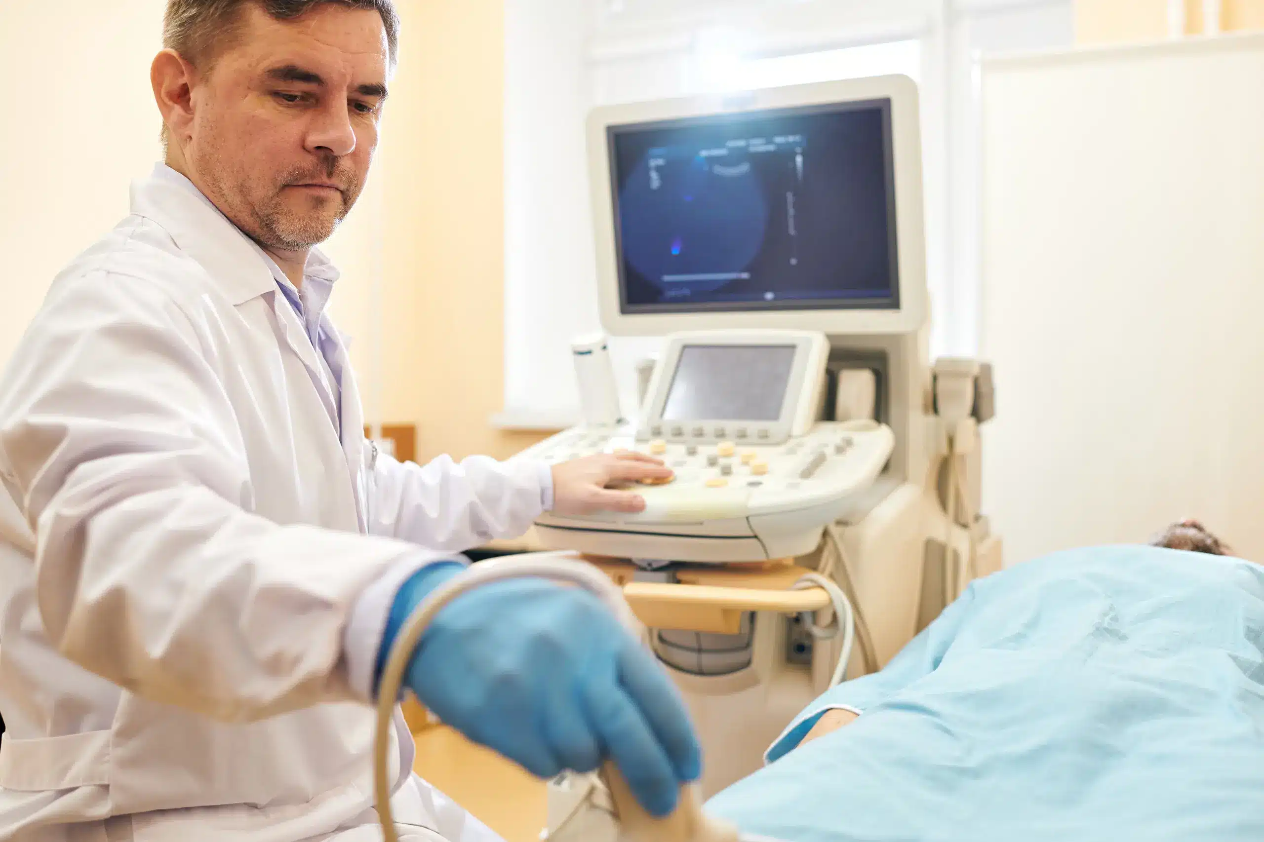
An endocavity ultrasound is one technique where an ultrasound transducer has been specially designed to be inserted through a natural orifice of the body. These are mainly the vagina, rectum, or the esophagus. Using these offers proximity to the organs to be imaged, thus improving the image quality and resolution. ECUS benefits, especially in structures that cannot be easily assessed by traditional abdominal ultrasound or by other imaging modalities.
The most extensive application of endocavity ultrasounds is in gynecology, and it is used mainly for assessing ovarian cysts, uterine fibroids, and the early complications of pregnancy. Highly detailed images of pelvic organs are produced through the TVUS, making it possible to aid in the diagnosis and treatment of a very wide range of conditions concerning reproductive health.
Lt represents a critical application of ultrasound, which is mainly conducted within urological sciences for pathology determination, for instance, tumors found within the prostate. It can be made as prostate biopsy guidance and may ultimately guide the treatment options for patients with PCa or other conditions.
Endoscopic Ultrasound, where an endoscope can obtain real-time ultrasound images of the esophagus and stomach. Gatrografin swallow is among the important diagnostic modalities that can detect a large number of gastrointestinal diseases like a tumor; cysts and other abnormalities.
Endocavity ultrasound is one of the types of ultrasound which are used in almost all fields of medical practice. Some of its applications, for example, include firewalls.
Obstetrically, TVUS is of excellent value since it follows up on the growth of the fetus together with complications related to pregnancy and assesses both the uterus and the ovaries. It diagnoses ectopic pregnancy and gestational trophoblastic syndromes around images.
TRUS is the reference standard when assessing prostate-related disorders. In this process, urologists are able to see the size and shape of the prostate gland and hypothesize abnormality. Besides this, TRUS is carried out to assess other pelvic structures including seminal vesicles and bladder.
EUS plays a vital role in the diagnosis and staging of gastrointestinal cancers. It provides nearly real-time views of the esophagus, the stomach, the pancreas, and its surrounding lymph nodes. Additionally, EUS results in fine-needle aspiration biopsies of pancreatic lesions that help establish a proper diagnosis.
In oncology, ECUS might be helpful in delineating the extent of tumors surrounding the natural cavities in the human body. For instance, EUS alone is used in the staging of oesophageal and gastric cancers; it enables clinicians to stage the degree of invasion and assists in the determination of nodal involvement.
Although it is not very frequently used, a cardiac environment could also make use of an endocavity ultrasound. For instance, TEE makes the anatomy of structures in the heart well visualized and hence would allow clinicians to achieve all the critical detections like valvular heart disease or cardiac masses.
Among the many benefits, one of the most promising advantages of the application of ECUS is the quality that it reaches in the image provided. By placing the transducer more proximal to the target organ, the clinicians will be able to have a much clearer view with finer details. Since the output would be very accurate, the diagnosis would get very precise.
Endocavity ultrasound will provide real-time imaging. This may make it possible for clinicians to capture dynamic processes like flows of blood or movement of organs. It is also one of those situations that require accuracy and, hence, much useful in such cases for guided biopsies.
ECUS is comparatively less invasive compared with others. It is least discomforting to the patient. The usage of a lubricant and also the transducer is so small that this makes the patient as comfortable as possible in the examinations.
Endocavity ultrasound can easily be modified for different applications among many specialties, thereby making the modality highly versatile in modern diagnostic imaging. With its ability to deliver significant information on anatomical regions, the scope of its applicability in clinical practice is inevitable.
After highlighting several advantages of endo cavity ultrasound, some significant limitations are as follows.
The quality of images obtained by endocavity ultrasound is significantly dependent on the experience and expertise of the practitioner. Variations in technique lead to variations in accuracy, thereby increasing the importance of training.
General In general, endocavity ultrasound provides only a relatively very limited field of view in contrast with other imaging techniques like CT or MRI. It can therefore not be used for surveying large areas or structures inaccessible with endocavity techniques.
Most patients can tolerate the procedure well; however, some may feel anxious or uncomfortable during the procedure. Effective communication and reassurance by healthcare workers may improve patient acceptance.
An infection may always be a risk with any procedure that requires insertion into a natural cavity. Proper hygiene and sterilization are therefore indispensable to minimize such risks.
With a push through technology and technical advances, prospects in the future for endocavity ultrasound look bright. It can be visualized that newer 3D and 4D ultrasound imaging would take diagnostic capabilities in ECUS to new heights by providing much more anatomical detail and, hence better assessments of complex structures.
Indeed, the future of ultrasound imaging will carry another added benefit by bringing artificial intelligence into the detection and analysis of various pathologies. Integrating AI into ultrasound imaging may offer more precise results for higher diagnostic detection. Also, images can be analyzed with AI algorithms in terms of improving efficiency and accuracy in the analysis of abnormalities.
It has thus emerged as a major addition to modern diagnostic imaging. Besides the many advantages of producing high-resolution images of internal anatomy, and versatility in a number of specialties, this has made ECUS a gold standard tool for the clinician.
Applications and benefits from ECUS are likely to extend even further and impact more strongly on patient care as this technology continues to advance.
For any other information or simply to learn more about endocavity ultrasound and our broader services for diagnostic imaging, contact Lincs Health (Pk). Our staff will be pleased to care for all your health care needs. If you want to know anything else related to endocavity ultrasound or diagnostic imaging use, or if you have any queries, kindly reach out to us at Lincs Health (Pk). Our team and we ensure that only the best solutions regarding health are presented to you, as per your needs.
A Specialized Radiology Center in Lahore is Providing Body , MSK, OBS Gynea TVS, Endocavity & Doppler Under Supervision of UK Trained Consultant in Lahore.
©2025, EBTECHSOL, All Rights Reserved.